Seeing Your Future Self with 3D Imaging
Modern aesthetic consultations now use advanced 3D imaging to transform how patients visualize potential surgical outcomes. Unlike traditional 2D photography, which flattens facial features and obscures depth, 3D technology creates highly accurate models from hundreds of data points. This precision allows patients to better understand their anatomy and align their goals with achievable results.
The scanning process is quick, non-invasive, and typically takes less than a minute. At drmmacdonald.com, we use these interactive models to bridge the communication gap, replacing subjective descriptions with a shared visual roadmap. By viewing potential changes from multiple angles, patients can make informed decisions with greater confidence.
Research highlights that these tools are essential for managing expectations before surgery. As AI-assisted simulations continue to evolve, they provide an increasingly personalized way to plan treatments that balance individual anatomy with desired aesthetic harmony.
The Role of 3D Imaging in Aesthetic Medicine
Unlike traditional 2D photography that flattens features and obscures critical contour depth, 3D imaging allows for a multi-perspective analysis of facial and body anatomy. Specialized systems such as VECTRA or Crisalix utilize photogrammetry and multi-camera arrays to capture hundreds of data points, creating detailed volumetric models with millimeter-level precision.
The capture process is efficient, often lasting less than a minute, and remains entirely non-invasive without the use of radiation. This documentation provides a visual roadmap for customized care, helping surgeons and patients align on achievable goals based on real anatomical data rather than idealized digital standards. At drmmacdonald.com, our approach integrates this technology to prioritize patient understanding and clinical accuracy.
While platforms like Aura provide powerful visualization, these simulations are predictive tools meant to guide planning rather than serve as absolute guarantees of surgical outcomes. As highlighted in The PMFA Journal, the true value lies in co-creating a treatment plan that respects the patient's unique tissue behavior and anatomical constraints. By transforming abstract descriptions into concrete visual data, this technology improves clarity and confidence across every stage of the aesthetic experience.
What to Expect During a 3D-Enhanced Consultation
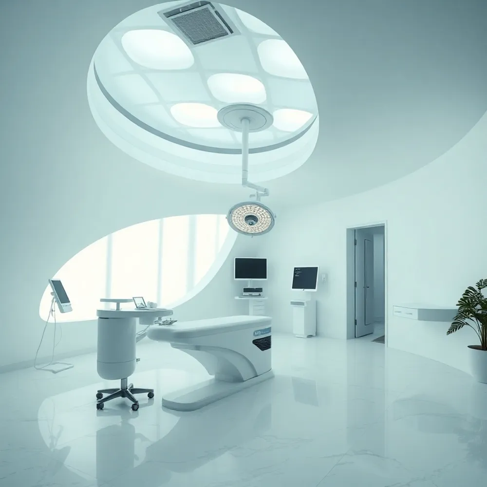
![]() Your visit to drmmacdonald.com begins with a thorough check-in and medical history review to ensure your safety and eligibility. Once this preparation is complete, you will meet with your surgeon to discuss your aesthetic goals and examine your concerns using 3D imaging. Unlike traditional consultations where surgeons must rely on verbal descriptions, this technology provides an objective visual baseline that helps clarify your unique anatomy.
Your visit to drmmacdonald.com begins with a thorough check-in and medical history review to ensure your safety and eligibility. Once this preparation is complete, you will meet with your surgeon to discuss your aesthetic goals and examine your concerns using 3D imaging. Unlike traditional consultations where surgeons must rely on verbal descriptions, this technology provides an objective visual baseline that helps clarify your unique anatomy.
The imaging session adds approximately 15 to 30 minutes to your visit, bringing the total appointment time to roughly one or two hours. To ensure the most accurate data, please arrive without heavy makeup for facial procedures or wearing form-fitting, dark clothing for body-focused assessments. This precision allows our team to capture hundreds of data points, creating a high-fidelity digital model that grounds our treatment planning in reality rather than subjective interpretation.
During your session, your surgeon will use the resulting 3D model to co-create a treatment plan tailored to your specific facial harmony. This collaborative process helps you visualize how different procedures may address your concerns, such as understanding why certain areas of the face may benefit from support rather than direct filling. By seeing these anatomical relationships in three dimensions, you can better understand how procedures may achieve natural-looking outcomes that align with your goals.
What should I expect during a plastic surgery consultation?
A plastic surgery consultation is a comprehensive, two-way dialogue designed to help you determine if a procedure is the right choice for your unique needs. You can typically expect the appointment to last between one and two hours, beginning with a check-in process and a review of your medical history by a medical assistant. You will then meet with your surgeon to discuss your aesthetic goals, areas of concern, and personal expectations to ensure you have a clear understanding of potential results. This session is the ideal time to ask detailed questions regarding the procedure, recovery process, and safety protocols to ensure you feel fully informed and confident in your decision. Ultimately, the consultation serves as a collaborative, professional environment where we prioritize your comfort, safety, and personalized care.
Clinical Photography vs. 3D Imaging
Clinical photography aims to accurately document a patient's physical state under consistent, standardized conditions. Unlike portrait photography, which seeks to aestheticize or alter the subject, clinical photography requires rigorous consistency to provide an objective baseline for pre-operative planning and post-operative progress tracking.
While traditional photography provides a necessary historical record, it flattens facial anatomy and obscures vital contour details. Modern 3D imaging bridges this gap by capturing hundreds of data points to generate volumetric models with millimeter-level precision. This approach allows surgeons to evaluate depth and proportions that remain invisible in 2D imagery.
To ensure reliable anatomical comparisons, 3D systems utilize standardized lighting and double-flash technology. At drmmacdonald.com, these tools provide a multi-perspective view, allowing for real-time simulations that help patients visualize potential surgical outcomes. This offers an interactive digital roadmap that clarifies goals before any intervention occurs.
Evaluating Facial Balance and Symmetry
![]() Professionals often refer to the Rule of Thirds, which divides the face into three equal segments, from the hairline to the brow, from the brow to the base of the nose, and from the base of the nose to the chin. While individual features vary, this framework serves as a guide for understanding facial proportions and balance during aesthetic planning. At drmmacdonald.com, practitioners pair these classic aesthetic principles with advanced 3D imaging to move beyond static 2D observations.
Professionals often refer to the Rule of Thirds, which divides the face into three equal segments, from the hairline to the brow, from the brow to the base of the nose, and from the base of the nose to the chin. While individual features vary, this framework serves as a guide for understanding facial proportions and balance during aesthetic planning. At drmmacdonald.com, practitioners pair these classic aesthetic principles with advanced 3D imaging to move beyond static 2D observations.
While some platforms provide generalized digital renderings, 3D imaging adds precision by capturing submillimeter measurements of symmetry and proportions across your unique anatomy. This technology allows surgeons to analyze the underlying bone structure, soft tissue, and aging patterns in detail. Because these digital models can be rotated and zoomed, you and your surgeon can perform a comprehensive 360-degree assessment of your facial harmony.
Using this high-fidelity data, surgeons can bridge the gap between your expectations and anatomical reality. This clinical approach helps ensure that any proposed plan, whether surgical or non-surgical, respects the natural balance of your features rather than forcing an idealized, one-size-fits-all standard.
The 3D Skin Analysis Experience
![]() What is involved in a 3D skin analysis? A 3D skin analysis typically utilizes advanced multi-spectral imaging to capture comprehensive data both on and beneath the skin's surface. During the session, the system scans the complexion to identify specific concerns such as textural variations, sun damage, pore size, and signs of aging that may not be apparent to the naked eye.
What is involved in a 3D skin analysis? A 3D skin analysis typically utilizes advanced multi-spectral imaging to capture comprehensive data both on and beneath the skin's surface. During the session, the system scans the complexion to identify specific concerns such as textural variations, sun damage, pore size, and signs of aging that may not be apparent to the naked eye.
This non-invasive process creates a detailed visual profile of your skin health. By establishing a clear, quantifiable baseline, this technology allows our specialists at drmmacdonald.com to track improvements over time with objective data.
Unlike generalized assessments, this 3D imaging methodology enables our team to customize highly personalized non-surgical rejuvenation treatments tailored to your unique anatomical needs. This diagnostic edge ensures you approach your treatment plan with greater precision and confidence.
Visualizing Outcomes for Common Facial Procedures
3D imaging brings a new level of clarity to consultations for rhinoplasty, facelifts, dermal fillers, laser peels, and chin or cheek implants. Instead of relying on static before-and-after photos of other patients, you see simulations tailored to your own anatomy. This personalized preview helps both you and your surgeon decide on the most appropriate approach before entering the operating room.
Rhinoplasty: High Accuracy with Predictable Structures
Rhinoplasty is considered one of the most accurate procedures for 3D simulation because the nose's underlying bone and cartilage structures are well defined. Surgeons can manipulate the digital model to show changes in bridge height, tip rotation, and nostril width with a high degree of reliability. The simulation serves as a shared visual roadmap — you can see how potential adjustments would affect overall facial balance before any incisions are made.
Facelifts and Soft Tissue Procedures
For facelifts and other soft tissue procedures, 3D imaging provides a useful approximation of how lifting and tightening will reshape the jawline, cheeks, and neck. Because soft tissues have more biological variability than cartilage or bone, these simulations are best viewed as strong visual guides rather than exact promises. They help clarify what is realistically achievable given your skin elasticity and healing patterns.
Dermal Fillers: Restoring Volume Naturally
Dermal filler simulations let you see how volume restoration in areas like the cheeks, nasolabial folds, or lips will change your facial proportions. The 3D model highlights how subtle adjustments to contour can restore balance without looking overdone. You can compare different levels of enhancement side by side to find a result that matches your aesthetic goals.
Intraoperative Use of 3D Models
Some surgeons take 3D imaging beyond the consultation room by projecting it onto a screen during surgery as an augmented reality guide. This overlay helps them stay aligned with the planned contours when reshaping bone or repositioning soft tissue. At drmmacdonald.com, we integrate this data into our surgical planning to support consistent, reproducible outcomes.
Setting Realistic Expectations and Building Confidence
3D imaging transforms the preoperative phase by replacing abstract concepts with a clear, anatomical roadmap. At drmmacdonald.com, this precision helps bridge the divide between a patient's vision and what individual tissue dynamics can realistically achieve.
These simulations function as predictive tools rather than absolute guarantees. While surgeons can manipulate models to demonstrate potential outcomes, factors like natural healing, skin elasticity, and underlying bone structure mean final results will always be unique to the person. This clarity is vital, as research indicates that setting honest, evidence-based goals early on significantly boosts patient satisfaction.
Side-by-side comparisons of current features against projected results provide the visual documentation required to make an informed decision. By reviewing these renderings, patients gain the confidence to proceed or identify when a specific request may fall outside the bounds of their anatomy. This proactive education is why practices using these digital platforms often see higher conversion rates of consultations into successful, customized surgical plans.
Emerging Trends: The Future of 3D Imaging and AI
The 2026 aesthetic landscape reflects an intense preference for natural-looking, sustainable results. Surgeons now move beyond broad interventions, focusing on personalized hybrid treatment plans that blend surgical precision with non-surgical, regenerative modalities. By prioritizing individual facial harmony, practitioners use advanced imaging to ensure that every correction complements a patient's unique bone structure and aging pattern.
What are the emerging trends in plastic surgery for 2026?
Regenerative medicine today relies on a patient’s own biological resources, such as fat grafting, to restore volume rather than defaulting to synthetic implants. The surge in patients utilizing GLP-1 medications has further altered the current demand, necessitating innovative body contouring techniques and specialized protocols for facial volume restoration. To remain agile, aesthetic surgery incorporates AI-driven analytics with real-time 3D scans to predict how tissues will respond to specific volume adjustments before the surgeon makes a single incision.
High-fidelity visualization is no longer limited to screen-based simulations. Innovations now allow for the creation of 3D-printed surgical guides and physical masks derived from scans, offering a tangible roadmap for highly complex facial procedures. While competitors may rely on basic photo-morphing, drmmacdonald.com integrates these immersive technologies to map out every anatomical nuance. This precision enables surgeons to execute procedures with millimeter-level accuracy, ensuring the final result matches the patient's goal for a refreshed, balanced, and rejuvenated look.
Empowering Your Aesthetic Journey with 3D Vision
3D imaging fundamentally transforms the consultation process by replacing abstract discussions with concrete, data-driven visualization. At drmmacdonald.com, we leverage these advanced models to foster a transparent dialogue, ensuring you understand exactly how proposed techniques align with your unique facial anatomy. While standard morphing tools exist elsewhere, our approach prioritizes anatomical precision over idealized digital standards, providing a reliable roadmap for your results.
- Enhanced communication that bridges the gap between surgical intent and patient understanding.
- Personalized treatment planning rooted in your specific measurements rather than generalized galleries.
- Clearer expectations regarding what is surgically achievable, promoting long-term confidence.
Technology is merely a support system. The true value emerges when these high-resolution simulations are interpreted by an experienced, board-certified surgeon who can translate digital projections into physical reality. As aesthetic medicine continues to evolve toward more personalized, AI-integrated care, utilizing these tools early in your planning phase ensures you remain an active, informed participant in your transformation. We invite you to book a consultation to experience how this technology helps illuminate your path to natural, harmonious results.